Products in stock in the United States can be delivered within 2 days.
 +1-2404726069 (U.S.) +86-027-65528241 (Outside U.S.)
  overseas@yeasen.com shop website:https://yeabio.com
 0
0MycAway™ Treatment (1000×) - Mycoplasma Elimination Reagent

Description
In the process of cell cultures, regular detection can effectively avoid mycoplasma infection, but if the cells have been infected with mycoplasma, they need to be treated in time. The best treatment is to discard the cells after high-pressure sterilization to avoid contaminating other clean cell lines. If contaminated cells are precious, mycoplasma infection must be removed. Because mycoplasma lacks the cell wall, conventional antibiotics are generally ineffective. This product is an improved mixed preparation specially used for mycoplasma elimination. It can achieve a good mycoplasma elimination effect by inhibiting the synthesis of related proteins and is not harmful to cells. Minimize the loss of your precious cells due to mycoplasma infection.
Feature
- Ultra-low Toxicity: Only block the bacterial protein synthesis rather than animal cells
- Excellent Stability: Stored at -15℃ ~ -25℃ for 18 months
- Facilitated Operation: Just added into the culture medium
- Rapid Onset: Take effect in 3 days
- Wide Applicability: Effective against most mycoplasmas
Application
- Mycoplasma Elimination
Components
| Components No. | Name | 40607ES03 | 40607ES08 |
| 40607 | MycAway™ Treatment (1000×) - Mycoplasma Elimination Reagent | 1 mL | 5×1 mL |
Shipping and Storage
The product is shipped with ice packs and can be stored at -15℃ ~ -25℃ for 18 months. Please keep it away from light.
Figures
- The representative image of the cell before and after mycoplasma removal
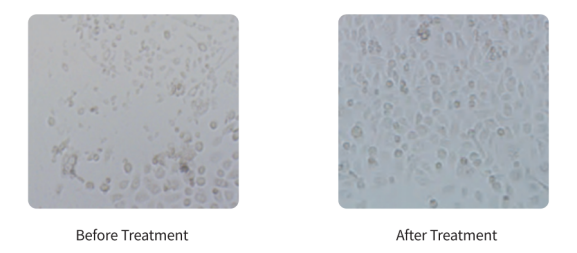
Figure 1. The representative image before and after mycoplasma removal by YEASEN MycAway™ Reagent
- The cell growth and viability were not affected by YEASEN MycAway™ Reagents
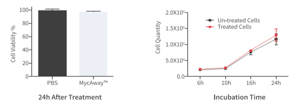
Figure 2:The effects of MycAway™ Reagents on cell growth and viability were detected.
The cell growth and viability were not affected by YEASEN MycAway™ Reagents.

[1] Sun C, Kang YF, Liu YT, et al. Parallel profiling of antigenicity alteration and immune escape of SARS-CoV-2 Omicron and other variants. Signal Transduct Target Ther. 2022;7(1):42. Published 2022 Feb 8. doi:10.1038/s41392-022-00910-6(IF:18.187)
[2] Malbec L, Zhang T, Chen YS, et al. Dynamic methylome of internal mRNA N<sup>7</sup>-methylguanosine and its regulatory role in translation. Cell Res. 2019;29(11):927-941. doi:10.1038/s41422-019-0230-z(IF:17.848)
[3] Wang Y, Wu T, Tang M. Ambient particulate matter triggers dysfunction of subcellular structures and endothelial cell apoptosis through disruption of redox equilibrium and calcium homeostasis [published correction appears in J Hazard Mater. 2021 Jan 15;402:123316]. J Hazard Mater. 2020;394:122439. doi:10.1016/j.jhazmat.2020.122439(IF:9.038)
[4] Wang Y, Liu N, Huang X, et al. Atmospheric particulate matter impedes autophagic flux by impairing lysosomal milieu and integrity in human umbilical vein endothelial cells (HUVECs). Sci Total Environ. 2021;761:143290. doi:10.1016/j.scitotenv.2020.143290(IF:6.551)
[5] Tang C, Wang X, Ji C, et al. The Role of miR-640: A Potential Suppressor in Breast Cancer via Wnt7b/β-catenin Signaling Pathway. Front Oncol. 2021;11:645682. Published 2021 Apr 12. doi:10.3389/fonc.2021.645682(IF:6.244)
[6] Wang Y, Tang M. PM2.5 induces autophagy and apoptosis through endoplasmic reticulum stress in human endothelial cells. Sci Total Environ. 2020;710:136397. doi:10.1016/j.scitotenv.2019.136397(IF:5.589)
Catalog No.:*
Name*
phone Number:*
Lot:*
Email*
Country:*
Company/Institute:*
