 +1-2404726069 (U.S.)
 0
0Apoptosis generally refers to a kind of programmed cell death that occurs through the regulation of intracellular genes and their products during the development of cells or under the action of some factors. Apoptosis plays an important role in embryonic development and morphogenesis, the stability of normal cell populations in tissues, the body's defense and immune response, cell damage and aging caused by disease or poisoning, and the occurrence and progression of tumors, and has potential therapeutic significance.
The events in the apoptotic pathway occur in a time sequence, that is, the events occur in sequence, and eventually lead to the emergence of apoptotic bodies, with the occurrence of apoptosis.
Its typical characteristics are as follows:
1. Early apoptosis: cell membrane structure changes, phosphatidylserine eversion;
2. Early and middle stage apoptosis: the density of cytoplasm increased, the mitochondrial membrane potential disappeared, the permeability changed, and cytochrome C was released into the cytoplasm;
3. Late apoptosis: apoptosis signal transduction;
4. Late apoptosis: DNA degraded to 180~200bp fragment size.
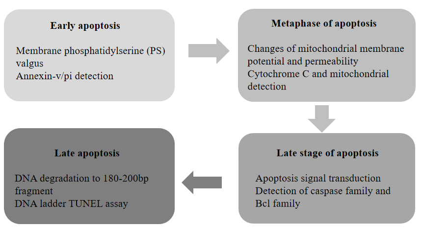
Figure 1: Occurrence and detection of sequential events in the apoptosis pathway
1. Early apoptosis: Annexin-V/PI double staining (detection of PS on the extracellular membrane)
In normal living cells, phosphatidylserine (PS) is located on the inner side of the cell membrane. When apoptosis occurs, the cell membrane changes, and PS flips from the inner surface of the cell membrane to the outer surface of the cell membrane. Annexin-V is a Ca2+ dependent phospholipid binding protein with a molecular weight of 35-36 KD, which can bind with PS with high affinity. Using Annexin-V labeled with fluorescein (FITC, Alexa fluor488, etc.) as a probe, apoptotic cells and living cells can be identified by flow cytometry or fluorescence microscope.
PS of necrotic cells will also turn over from the inner surface of cell membrane to the outer surface of cell membrane. Annexin-V can also recognize PS on the surface of necrotic cells, so Annexin-V cannot distinguish necrotic cells from apoptotic cells. In addition, propidine iodide (PI) is a nucleic acid dye, which can bind to DNA in cells, and it cannot penetrate the complete cell membrane. The cell membrane of early apoptotic cells and living cells is still intact, and PI dye cannot enter the cell freely through the cell membrane to bind with DNA, so PI dye cannot label apoptotic cells and living cells, but PI dye can bind with DNA in the cell through the cell membrane of necrotic cells. PI dye in the dead cells will emit red fluorescence after being excited by 488nm laser, and will be received by the corresponding channel. Therefore, Annexin V and PI can be used together to distinguish living cells, early apoptotic cells and necrotic cells.
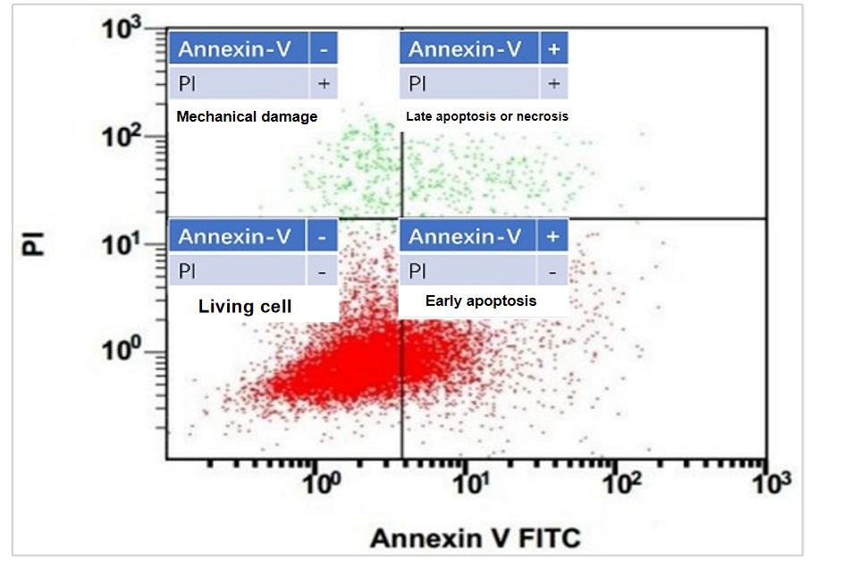
Figure 2: Analysis of flow cytometry results after annexin-v/pi double staining
Ordering products:
| Product information | Cat number | Specifications |
| Annexin V-FITC/PI Apoptosis Detection Kit | 40302ES20 | 20T |
| 40302ES50 | 50T | |
| 40302ES60 | 100T | |
| Annexin V-EGFP/PI Apoptosis Detection Kit | 40303ES20 | 20T |
| 40303ES50 | 50T | |
| 40303ES60 | 100T | |
| Annexin V-Alexa Fluor 647/PI Apoptosis Detection Kit | 40304ES20 | 20T |
| 40304ES50 | 50T | |
| 40304ES60 | 100T | |
| Annexin V-Alexa Fluor 488/PI Apoptosis Detection Kit | 40305ES20 | 20T |
| 40305ES50 | 50T | |
| 40305ES60 | 100T | |
| Annexin V-PE/7-AAD Apoptosis Detection Kit (Inquire) | 40310ES20 | 20T |
| 40310ES50 | 50T | |
| 40310ES60 | 100T |
2. Early apoptosis: JC-1 staining (detection of mitochondrial membrane potential changes)
The process of apoptosis is often accompanied by the destruction of mitochondrial transmembrane potential, which is widely considered to be one of the earliest events in the process of the apoptosis cascade. It occurs before the appearance of the characteristics of nuclear apoptosis (chromatin concentration, DNA breakage). Once the mitochondrial transmembrane potential collapses, cell apoptosis is irreversible. The existence of mitochondrial transmembrane potential enables some lipophilic cationic fluorescent dyes such as rhodamine 123, JC-1, JC-10 to bind to the mitochondrial matrix. The increase or decrease of their fluorescence indicates the increase or decrease of the electronegativity of the mitochondrial inner membrane.
JC-1 is widely used to detect mitochondrial membrane potential △ΨM, showing potential-dependent accumulation in mitochondria. In normal mitochondria, JC-1 aggregates in the mitochondrial matrix to form a polymer, which emits strong red fluorescence (ex=585 nm, em=590 nm); In apoptotic cells, mitochondrial transmembrane potential depolarized, JC-1 was released from mitochondria, the concentration decreased, and reversed to the monomer form emitting green fluorescence. Therefore, the change of color directly reflects the change in mitochondrial membrane potential. The degree of mitochondrial depolarization can also be measured by the ratio of red/green fluorescence intensity. JC-1 detection is a common method.
| Product name | Cat number | Specifications |
| JC-1 fluorescent probe (Inquire) | 40705ES03 | 1mg |
| 40705ES08 | 5mg | |
| JC-10 mitochondrial membrane potential fluorescent probe (Inquire) | 40707ES03 | 1mg |
| 40707ES08 | 5mg | |
| JC-1 Mitochondrial Membrane Potential Assay Kit (Inquire) | 40706ES60 | 100T |
3. Late apoptosis: TUNEL method (DNA fragment in situ labeling method)
At the late stage of apoptosis, a large number of sticky 3-OH terminals are produced due to double strand breaks or single strand breaks of chromosomal DNA. Under the action of deoxyribonucleotide terminal transferase (TdT), luciferase labeled dUTP can be bound to the 3-terminal of DNA, so that apoptotic cells can be detected, such methods are called terminal deoxynucleotidyl transferase mediated dUTP nick end labeling (TUNEL). Because normal or proliferating cells have almost no DNA breaks, there is no 3-OH formation and few can be stained. TUNEL is actually a research method combining molecular biology and morphology. In situ staining of a complete single apoptotic nucleus or apoptotic body can accurately reflect the typical biochemical and morphological characteristics of apoptosis. It can be used to determine the cell morphology of paraffin embedded tissue sections, frozen tissue sections, cultured cells and cells isolated from tissues, and can detect a very small number of apoptotic cells, Therefore, it is widely used in the study of apoptosis.
| Product information | Cat number | Specifications |
| TUNEL Apoptosis Detection Kit (FITC) | 40306ES20 | 20T |
| 40306ES60 | 100T | |
| TUNEL Apoptosis Detection kit ( Alexa Fluor 488) | 40307ES20 | 20T |
| 40307ES60 | 100T | |
| TUNEL Apoptosis Detection kit (Alexa Fluor 640) | 40308ES20 | 20T |
| 40308ES60 | 100T |
Related products:
|
Product information |
Cat number |
Specifications |
|
Cell Cycle and Apoptosis Analysis Kit (Inquire) |
40301ES50 |
50T |
|
40301ES60 |
100T |
|
|
PI (Propidium iodide) (Inquire) |
40711ES10 |
10mg |
|
40711ES60 |
100mg |
|
|
Rhodamine 123 (Inquire) |
40712ES08 |
5mg |
|
DAPI (Inquire) |
40727ES10 |
10mg |
|
DAPI Stain Solution (Inquire) |
40728ES10 |
10ml |
|
40728ES50 |
50ml |
|
|
Hoechst 33258 (Inquire) |
40729ES25 |
25mg |
|
Hoechst 33258 Stain Solution (Inquire) |
40730ES10 |
10ml |
|
40730ES50 |
50ml |
|
|
Hoechst 33342 (Inquire) |
40731ES25 |
25mg |
|
Hoechst 33342 Stain Solution (Inquire) |
40732ES10 |
10ml |
|
40732ES50 |
50ml |
