Products in stock in the United States can be delivered within 2 days.
 +1-2404726069 (U.S.) +86-027-65528241 (Outside U.S.)
  overseas@yeasen.com shop website:https://yeabio.com
 0
0CHO Host Cell DNA Residue Detection Kit

Product Description
CHO Host Cell DNA Residue Detection Kit is used for the quantitative analysis of CHO host cell DNA residuce in intermediate samples, semi-finished and finished products of various biological products.
This kit adopts Taqman fluorescent probe and the polymerase chain reaction (PCR) method, which has fg level minimum detection limit and can specifically and quickly detect the residual CHO cell DNA. The kit needs to be used together with the the Residual DNA Sample Preparation Kit (Cat# 18461ES).
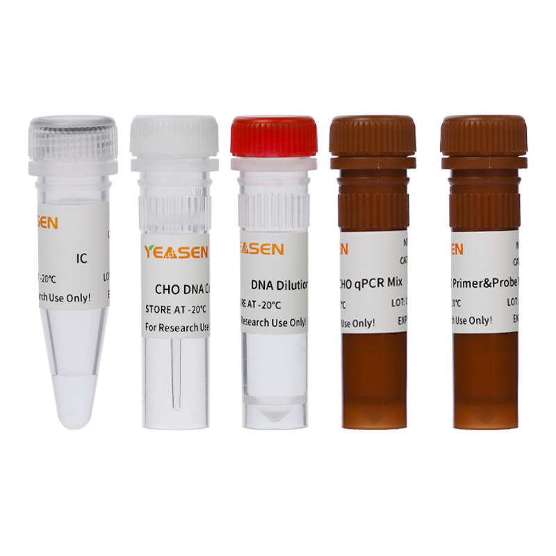
Product Components
|
Category |
Components No. |
Components Name |
Cat#/Size |
|
|
41301ES50 (50T) |
41301ES60 (100T) |
|||
|
Part I |
41301-A |
CHO qPCR mix |
1 mL |
2 mL |
|
41301-B |
DNA Dilution Buffer |
2×2 mL |
4×2 mL |
|
|
Part Ⅱ |
41301-C |
CHO DNA Control (30 ng/μL) |
25 μL |
50 μL |
[Notes]: This kit does not contain ROX reference dye. If you need to add ROX reference dye to the Real Time PCR thermal cycler you are currently using, it is recommended that you purchase 50× ROX Reference Dye (Cat#10200ES) from our company.
Shipping and Storage
1. Part I is shipped on dry ice and stored at -20°C for 2 year
2. Part II is shipped on dry ice and stored at -80°C for 2 year.
3. After receiving the goods, please check whether the two components of Part I and Part II are complete, and store them in the corresponding storage temperature immediately.
Cautions
1. Please read this manual carefully before using this reagent, and the experiment should be standardized, including sample handling, reaction system preparation and sample addition.
2. Adding samples and preparing solutions is best done on ice.
3. Ensure that each component is fully vortexized and centrifuged at low speed before use.
4. For your safety and health, please wear lab coats and disposable gloves for operation.
5. This product is for research use ONLY!
Applicable instrument models
Include but not limited to:
Bio-Rad: CFX96 Optic Module.
Thermo Scientific: ABI 7500; ABI Quant Studio 5; ABI Step OnePlus.
Using Instruction
1. CHO DNA Standard dilution and Standard curve preparation
The CHO DNA Control was gradient diluted using the DNA Dilution Buffer provided in the kit, and the dilution concentration is 300 pg/μL, 30 pg/μL, 3 pg/μL, 300 fg/μL, 30 fg/μL, 3 fg/μL.
See detailed instructions below:
1.1 Thaw the CHO DNA control and DNA dilution buffer on ice. After completely thawed, vortex gently to mix, and centrifuge at low speed for 10 secs.
1.2 Take out six clean 1.5 mL tubes, marked with 3 ng/μL, ①, ②, ③, ④, ⑤, ⑥.
1.3 Add 90 μL DNA dilution buffer and 10 μL CHO DNA Control to the 1.5 mL microfuge tube labeled 3 ng/μL, dilute to 3 ng/μL. Mix and then centrifuge for 10 secs. Subpackage the diluted DNA standard and it can be stored in the short term (no more than 3 months) at -80°C.Please avoid repeated freeze-thaw.
1.4 Add 90 μL DNA dilution buffer into other tubes, then follow the below procedure for the serial dilutions.
|
Tube |
Dilution Ratio |
Standard concentration |
|
① |
10 μL 3 ng/μL+90 μL DNA Dilution Buffer |
300 pg/μL |
|
② |
10 μL ①+90 μL DNA Dilution Buffer |
30 pg/μL |
|
③ |
10 μL ②+90 μL DNA Dilution Buffer |
3 pg/μL |
|
④ |
10 μL ③+90 μL DNA Dilution Buffer |
300 fg/μL |
|
⑤ |
10 μL ④+90 μL DNA Dilution Buffer |
30 fg/μL |
|
⑥ |
10 μL ⑤+90 μL DNA Dilution Buffer |
3 fg/μL |
[Notes]:
1. Three replicate wells are required for each concentration.The detection range is 3 fg/μL-300pg/μL and this range can be expanded if required.
2. To reduce the number of repeat freeze-thaw and avoid contamination, it is recommended to store the DNA control in aliquots at -80°C for the first time.
3. Once thawed, DNA dilution buffer could be stored at 2-8°C for 7 days, if not used for a long time, please store at -20°C.
4. Make sure the template is completely mixed, gently shake the mixture for 15 secs to 1 min for each gradient dilution.
2. Extraction Recovery Control (ERC) preparation
Set the concentration of CHO DNA in ERC as needed (the ERC sample was prepared with 3 pg/μL DNA as an example), as follows:
2.1 Add 100 μL test sample into a clean 1.5 mL tube, then add 10 μL 3pg/μL CHO DNA Standard (③) and mix well, marked as ERC.
2.2 Perform the DNA extraction of ERC sample together with the test samples to prepare the purified ERC sample.
3. Positive Control Sample (PCS) preparation (Optional)
Set the concentration of CHO DNA in PCS as needed (the PCS was prepared with 3 pg/μL DNA as an example), as follows:
3.1 Add 100 μL 3 pg/μL CHO DNA Standard (③) into a clean 1.5 mL tube, then marked as PCS.
3.2 Perform the DNA extraction of SRC together with the test samples to prepare the purified PCS.
4. Negative Control Solution (NCS) preparation
Set the negative control in the experiment, the specific operation steps are as follows:
4.1 Add 100 μL sample matrix (or DNA dilution buffer) into a clean 1.5 mL tube, then marked as NCS.
4.2 Perform the DNA extraction of NCS sample together with the test samples to prepare the purified NCS sample.
5. No Template Control (NTC) preparation
Set the no template control in the experiment, the specific operation steps are as follows:
5.1 NTC requires no sample pretreatment, and can be configured at the stage of qPCR detection of residual DNA content.
5.2 The NTC sample in each tube or well is 20 μL CHO qPCR Mix + 20 μL DNA Dilution Buffer. It is recommended to configure three replicate wells.
6. PCR reaction system
|
Component |
Volume(μL) |
|
CHO qPCR mix |
20 |
|
DNA template |
20 |
|
Total volume |
40 |
[Notes]:
1. Calculate the total PCR reaction volume by the number of reactions: qPCR Mix =(the number of reactions+2) ×20 μL (including the losses of two reaction wells). More than three replicates for each sample are recommended in the experiment.
2. After capping the tube or sealing the plate, centrifuge the reaction tube or plate at low speed for 10 secs. After sufficient shaking and mixing for 5 secs, repeat centrifuge to collect the liquid from the lid or wall to the bottom. Avoid bubbles during operation.
See below table for the recommended Plate setup:
|
|
1 |
2 |
3 |
4 |
5 |
6 |
7 |
8 |
9 |
|
A |
NTC |
|
STD 1 |
STD 1 |
STD 1 |
|
TS 1 |
TS 1 |
TS 1 |
|
B |
NTC |
|
STD 2 |
STD 2 |
STD 2 |
|
TS 2 |
TS 2 |
TS 2 |
|
C |
NTC |
|
STD 3 |
STD 3 |
STD 3 |
|
TS 3 |
TS 3 |
TS 3 |
|
D |
NCS |
|
STD 4 |
STD 4 |
STD 4 |
|
|
|
|
|
E |
NCS |
|
STD 5 |
STD 5 |
STD 5 |
|
ERC 1 |
ERC 1 |
ERC 1 |
|
F |
NCS |
|
STD 6 |
STD 6 |
STD 6 |
|
ERC 2 |
ERC 2 |
ERC 2 |
|
G |
|
|
|
|
|
|
ERC 3 |
ERC 3 |
ERC 3 |
|
H |
|
|
|
|
|
|
PCS |
PCS |
PCS |
The plate layout includes: 1 NTC (no template control), 1 NCS (negative control solution), 6 STD (the standard curve of 6 standard concentrations), 3 TS (test samples), 3 ERC (extraction recovery control), 1 PCS (positive control sample). Three replicate wells for each sample.
7. Setup guidelines for a PCR Instrument (2-step method)
The following instructions apply only to Bio-rad CFX 96 qPCR instrument (Software version 4.1.2433.1219). If you use a different instrument, refer to the applicable instrument guide for setup guidelines.
7.1 Generate a blank program, choose the template of absolute quantification;
7.2 Generate a detection probe and named as CHO-DNA, choose ‘user-define’ under ‘select run type‘ in the column of ‘run setup’, then set the program, plate layout and detection channels.
7.2.1 Click ‘Create New’ under ‘protocol’ to start the PCR program setting, then click ‘OK’ and save the program document.
7.2.2 Click ‘Next’ or ‘Plate’ and enter the plate layout interface, click ‘create new’, All Channels or SYBR/FAN Only can be choose in the ‘Scan Mode’. Chose the reaction box then click ‘select fluorophores’, chose FAM and click ‘OK’. Under ‘sample type’, chose unknow (or other sample type), choose the box on the right of ‘load’ in ‘target name’, and then set the ‘target name’ or not. Enter the sample names, biological groups according to the actual requirement or not. Then click ‘OK’ to save the final result.
7.2.3 Click ‘Next’ or ‘Start run’ to start the instrument
7.3 Set the amplification program: set the reaction volume as 40 μL.
7.3 Set the amplification program: set the reaction volume as 40 μL.
|
Cycle Step |
Temperature(℃) |
Time |
Cycles |
|
Initial denaturation |
95 |
5 mins |
1 |
|
Denaturation |
95 |
15 secs |
40 |
|
Annealing/Extension (Fluorescence collection) |
60 |
30 secs |
8. Analysis of qPCR results
8.1 After the program stopping, click the view/edit plate in the plate setup column in the upper right corner of the program, select the standard and choose ‘standard’ under the sample type column on the right side, set the repetition hole of each gradient, enter the concentration in ‘load concentration’ and press enter and finish setting the gradient of the standard in turn.
8.2 Click ‘OK’ to return to the initial screen and the standard curve will appear including the slope (Slope), intercept (Intercept), R², and amplification efficiency (Eff%). For a normal standard curve, R²>0.99; 90%≤Eff%≤110%.
8.3 Customers can set the programs according to their own needs and then the assay values of NTC, NCS, TS, ERC and PCS, the unit is fg/μL, and subsequently the units can be convert to pg/μL or pg/mL in the assay report.

Catalog No.:*
Name*
phone Number:*
Lot:*
Email*
Country:*
Company/Institute:*

